Explained
What is an OCT scan and what can it tell you?
Last updated: 20 August 2026
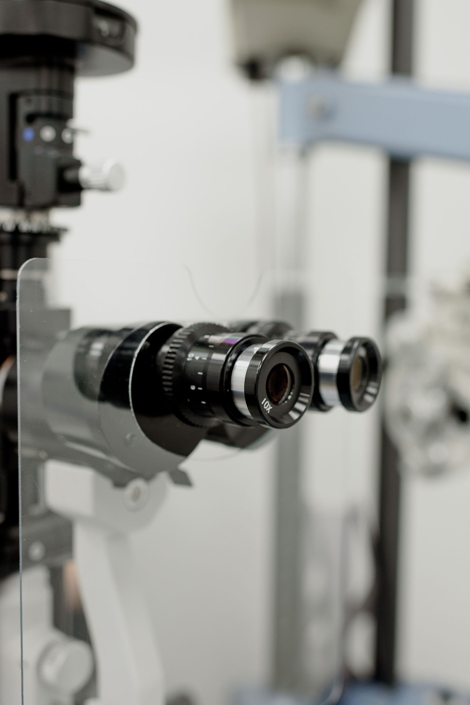
You encounter it ever more often at the optician, optometrist or ophthalmologist: the OCT scan. An impressive device, a cross-section of your retina in seconds — but what does such a scan actually say, and what doesn't it? A level-headed explanation.
In short
- OCT (optical coherence tomography) uses harmless light to image a cross-section of retina and optic nerve.
- The test is painless, touch-free and takes seconds per scan.
- Strong at: the macula (AMD, fluid), the nerve fibre layer (glaucoma), and tracking change over time.
- An OCT is an instrument, not a verdict: results gain meaning only combined with other examination.
⚠ Acute symptoms?Sudden vision loss, an eye injury, chemicals in the eye, severe pain, or new blind spots in your visual field? Don't wait for an online explanation — call your GP (huisarts), the out-of-hours GP service, or 112 immediately.
How does it work?
OCT stands for optical coherence tomography. The device sends a harmless beam of light into the eye and measures how the layers of the retina reflect it — comparable to ultrasound, but with light instead of sound. The result is a razor-sharp cross-section showing the individual layers of the retina, down to thousandths of a millimetre.
For you the test is simple: chin on the rest, look at a light, hold still for a few seconds. No contact, no radiation, usually no drops.
What does an OCT show well?
The scan excels in two places. The first is the macula, the centre of sharp vision. Fluid, swelling, a small hole, pulling scar tissue or the hallmarks of macular degeneration: an OCT often shows them earlier and more clearly than any other test. The second is the optic nerve and the nerve fibre layer around it. That layer's thickness is compared against a normative database — thinning can be an early sign of glaucoma, before anything is noticeable in the visual field.
At least as valuable as a single scan is the repeat scan: comparing measurements over months and years reveals whether something is stable or changing. For monitoring glaucoma and macular degeneration, that trend often matters more than the single snapshot.
What does an OCT not show?
An OCT is not a complete eye examination. The far edges of the retina fall outside the scanning field — a peripheral tear can remain invisible. It does not measure your prescription, nor your eye pressure, and it says little about cataract. A 'green' result within the normative range is reassuring but not a guarantee: some abnormalities require other tests.
The reverse also holds: a 'red' box in the report is not yet a diagnosis. The comparison against the database can be off because of a large or small optic nerve, a high prescription, or plain anatomical variation. Interpretation remains human work.
When is an OCT worthwhile?
With complaints about central vision (distortion, a spot in the middle), with elevated eye pressure or glaucoma in the family, for monitoring known macular or nerve conditions, and as a baseline against which later scans can be compared. Many opticians offer an OCT as part of an extended eye exam — a fine low-threshold first sieve, as long as it is clear that an abnormal scan calls for professional assessment.
What to do with a result that raises questions?
Ask for the image and the measurement report — you are entitled to them — and have someone explain what is abnormal and why it is or is not urgent. A second look at an existing scan is often more useful than immediately making a new one. And keep your reports: with OCT especially, the information lives in the trend.
Frequently asked questions
Is an OCT scan harmful or painful?
No. The device uses ordinary light (no X-rays), never touches the eye, and the test takes seconds. It can be repeated as often as needed.
Do I need dilating drops?
Usually not; modern devices scan through a normal pupil. With small pupils or cloudy media (such as cataract), drops can improve image quality.
My report shows red and yellow boxes. Is that serious?
Red and yellow mean: deviating from the database average. That can indicate a condition, but also harmless anatomical variation. Always have the report interpreted by whoever assesses the scan — the colour itself is not a diagnosis.
What does an OCT scan cost?
At many opticians it is part of a (free or paid) extended eye exam; in hospital it falls under the consultation. Ask beforehand — differences are large.
Sources and further reading
This article is general explanation, not medical advice about your situation. For acute symptoms: call your GP, the out-of-hours GP service, or 112.